Category:Magnetic resonance imaging
Jump to navigation
Jump to search
Deutsch: Magnetresonanztomographie.
non-destructive technique for imaging internal structures of objects or organisms | |||||
| Upload media | |||||
| Instance of | |||||
|---|---|---|---|---|---|
| Subclass of |
| ||||
| Based on | |||||
| |||||
Subcategories
This category has the following 36 subcategories, out of 36 total.
*
C
- Cine magnetic resonance imaging (40 F)
D
- Diffusion weighted imaging (3 F)
E
- Echo-planar imaging (2 F)
F
- Fossil MRI (12 F)
G
- Gradient echo (1 F)
H
- HARP (algorithm) (2 F)
I
- Inversion recovery MRI (2 F)
M
P
- Proton density imaging (1 F)
S
- Magnetic resonance imaging signs (14 F)
T
- Turbo spin echo (1 F)
Pages in category "Magnetic resonance imaging"
This category contains only the following page.
Media in category "Magnetic resonance imaging"
The following 200 files are in this category, out of 215 total.
(previous page) (next page)-
-2022-01-10 The Mammography, DEXA (osteoporosis diagnosis) and MRI Unit, Cromer Hospital.JPG 4,106 × 2,857; 1.89 MB
-
100325 RS88UH 11 qc.gif 480 × 480; 141 KB
-
202212 MRI Child.svg 512 × 512; 286 KB
-
202212 MRI Female.svg 512 × 512; 261 KB
-
202212 MRI Male.svg 512 × 512; 261 KB
-
3He MRI.jpg 291 × 323; 28 KB
-
872-4-8 (n).png 256 × 256; 114 KB
-
Ajmga62351-fig-0002-m.webp 500 × 263; 69 KB
-
Amyloid deposition diagnosis 01.png 1,200 × 1,077; 1.01 MB
-
Angio MR.jpg 589 × 586; 58 KB
-
Apple seed256.jpg 256 × 256; 19 KB
-
Arm mri.jpg 418 × 358; 94 KB
-
Artefact.jpg 2,825 × 2,757; 1,004 KB
-
Artefakt durch Zahnspange - MRT T2ax DWIax T1cor - 001.png 1,968 × 925; 464 KB
-
Beckenendlage MRT.jpg 1,074 × 1,110; 111 KB
-
Bilateral Sciatic Neurography.jpg 586 × 557; 212 KB
-
BIR-1 pulse for 90-degree and 45-degree flip-angles.png 1,278 × 743; 36 KB
-
Brain-Computer intrusive.jpg 400 × 600; 32 KB
-
BrainMRI3planes.gif 326 × 83; 1.91 MB
-
Bruker Biospec 94 30 MRI.jpg 6,936 × 9,248; 13.19 MB
-
Carotid MIP.jpg 652 × 644; 96 KB
-
Cervical MRI 120703 t2 me2d.png 474 × 339; 130 KB
-
Cochlea and semicirculars MRI 115700 MAIP cr.png 480 × 404; 120 KB
-
Colourised Axial MRI brain scan.webm 3.0 s, 1,080 × 1,080; 5.45 MB
-
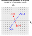
ConjugateSymmetryOfKspace.png 1,139 × 1,261; 129 KB
-
Connectivity matrix.jpg 852 × 807; 96 KB
-
Conventional MRI of the chest.jpg 289 × 333; 29 KB
-
Conventional MRI.jpg 307 × 269; 23 KB
-
Coronal Image of a TOFI and a Normal Control.jpg 720 × 960; 70 KB
-
CSF Flow vs Inspiration.png 695 × 520; 66 KB
-
CT and MRI of a red-eared slider (Trachemys scripta) - journal.pone.0017879.g001.png 2,009 × 2,941; 2.46 MB
-
Darmiyan Brain Map.png 1,186 × 1,127; 417 KB
-
DARPA Brain.jpg 800 × 486; 174 KB
-
De-Magnetresonanztomographie.ogg 3.5 s; 34 KB
-
Delayed (10 min ) Axial MR image IR.jpg 256 × 256; 40 KB
-
Doberman C6-C7 and C5-C6 traction responsive myelopathy A.jpg 409 × 268; 28 KB
-
Doberman C6-C7 and C5-C6 traction responsive myelopathy B.jpg 400 × 268; 22 KB
-
Doberman C6-C7 and C5-C6 traction responsive myelopathy.jpg 824 × 268; 50 KB
-
DTschematic.jpg 578 × 616; 44 KB
-
Ellipsoid eigenvalues14142.jpg 560 × 420; 20 KB
-
Ellipsoid2722.jpg 560 × 420; 21 KB
-
Ellipsoid888.jpg 560 × 420; 24 KB
-
EMI shielding faraday cage.jpg 1,944 × 2,592; 1.08 MB
-
EPI farsi.png 729 × 348; 45 KB
-
Fetal MRI.jpg 71 × 96; 2 KB
-
Four Wire.png 1,403 × 597; 52 KB
-
Gadoversetamide.png 830 × 335; 15 KB
-
Gd cosmos.jpg 1,800 × 1,800; 360 KB
-
GE table.jpg 619 × 417; 114 KB
-
GolaySet.png 487 × 630; 46 KB
-
GPUopen.JPG 3,648 × 2,736; 4.46 MB
-
Gpus .jpeg 866 × 1,155; 144 KB
-
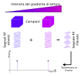
Gradiente de RMN.png 606 × 344; 36 KB
-
Haxby2001.jpg 763 × 500; 108 KB
-
Head mri animation.gif 610 × 256; 13.49 MB
-
Hierarchy animate.gif 600 × 184; 630 KB
-
Hyperpolarized MR.jpg 286 × 269; 21 KB
-
Idh1mut gbm.jpg 279 × 581; 72 KB
-
In vivo hyperpolarized carbon 13 MRI spectra.png 560 × 420; 29 KB
-
Inner ear Chochlea and vestibular system 115700 MAIP3 cr.png 480 × 394; 108 KB
-
IRM TEP acqusition protocols.png 1,522 × 585; 335 KB
-
Isointensitaet.JPG 506 × 802; 41 KB
-
IVIM1.jpg 270 × 400; 67 KB
-
Journal.pone.0057573.g005.png 909 × 588; 550 KB
-
K-space partial Fourier.JPG 415 × 372; 60 KB
-
Karman-Vortex-Realtime-MRI.ogv 20 s, 508 × 508; 1.31 MB
-
Kiwi 3d MRT projektion pls.gif 410 × 256; 9.46 MB
-
Kiwi 3d MRT projektion-endless.gif 410 × 256; 5.56 MB
-
Kiwi 3d MRT projektion.gif 256 × 410; 12.86 MB
-
Kspace farsi.png 546 × 316; 14 KB
-
Kspace multicoil.png 503 × 375; 56 KB
-
Kspace.png 566 × 516; 214 KB
-
Kspace2.png 242 × 240; 38 KB
-
Kspacetext.png 378 × 353; 107 KB
-
Larynx PDW nima.jpg 1,024 × 768; 137 KB
-
Lascar.jpg 581 × 288; 34 KB
-
LCIA Sign.jpg 2,048 × 1,536; 711 KB
-
LowContrast001.jpg 850 × 207; 41 KB
-
Lower extremity 111424 rgbca.png 550 × 248; 165 KB
-
Lower Extremity 111505 rgbca.png 537 × 241; 132 KB
-
Lower Limb MRL in healthy participant.tif 1,024 × 2,080; 8.22 MB
-
Mach mau.jpg 756 × 607; 77 KB
-
Mag suscp phantom.png 217 × 200; 26 KB
-
Magic angle.png 615 × 242; 13 KB
-
Magic angle.svg 600 × 300; 18 KB
-
Magnetic nanochain.jpg 3,607 × 2,403; 1.48 MB
-
Magnetic nanochains.jpg 3,607 × 2,403; 1.47 MB
-
Magnetic Resonance Imaging.png 1,904 × 1,516; 1.4 MB
-
Magnetic Resonance Research Centre West Cambridge.jpg 4,032 × 3,024; 2.22 MB
-
Maxillary sinus MRI.jpg 384 × 244; 25 KB
-
Maxwell pair.png 376 × 309; 19 KB
-
Media51b.webm 9.0 s, 696 × 720; 5.36 MB
-
Methods of imaging the lymphatic system.jpg 2,250 × 3,250; 754 KB
-
Motion.png 320 × 320; 91 KB
-
Motion2.png 320 × 320; 5 KB
-
MR CoA collaterals.png 599 × 452; 136 KB
-
MR obraz.jpg 1,245 × 1,107; 117 KB
-
MR-based attenuation correction.png 1,114 × 467; 278 KB
-
MRCP normal.tif 1,420 × 1,420; 674 KB
-
Mrfm.png 2,096 × 1,633; 1.51 MB
-
MRI Aliasing.jpg 256 × 168; 6 KB
-
MRI brain surface normal.jpg 691 × 664; 173 KB
-
Mri head 3dani 1 bionerd.gif 624 × 759; 3.86 MB
-
MRI Head Brain Normal.jpg 697 × 673; 196 KB
-
MRI Image of Human Head (94-087-1).jpeg 2,048 × 2,560; 277 KB
-
MRI knee abdonrmal.jpg 523 × 672; 126 KB
-
MRI of intestines.jpg 512 × 512; 94 KB
-
MRI of rib cage.jpg 512 × 512; 81 KB
-
MRI of torso.jpg 512 × 512; 100 KB
-
MRI Part 2.png 768 × 1,024; 540 KB
-
MRI Phase Encoding Principle.svg 420 × 154; 242 KB
-
MRI readout.svg 650 × 600; 34 KB
-
MRI RF shielding.jpg 1,944 × 2,592; 827 KB
-
MRI ringing.png 512 × 416; 71 KB
-
MRI Scan General Illustration.jpg 907 × 895; 288 KB
-
MRI Scanner at Narayana Multispeciality Hospital, Jaipur.jpg 4,496 × 3,000; 5 MB
-
Mri scanner schematic malayalam.png 1,178 × 1,082; 183 KB
-
MRI Scanner.jpg 3,120 × 4,160; 3.33 MB
-
MRI scanning of museum specimens - journal.pone.0034499.g002.png 2,073 × 2,778; 4.25 MB
-
MRI schema gradienti.svg 587 × 321; 15 KB
-
MRI selezione fetta.svg 950 × 400; 15 KB
-
MRI showing left frontal haemorrhage on SWI sequence.jpg 384 × 424; 80 KB
-
MRI spin echo.svg 628 × 294; 197 KB
-
MRI. elbow. ulnar collateral ligament.jpg 798 × 560; 108 KB
-
MRI. Intact inferior GHL.jpg 1,478 × 1,122; 222 KB
-
MRI. intact triceps tendon sagittal..jpg 1,478 × 1,122; 244 KB
-
MriDemoFl.gif 192 × 253; 571 KB
-
MRIGradientMalayalam.png 585 × 409; 43 KB
-
MriScanner.gif 442 × 280; 7 KB
-
MRN of ulnar nerve branches in Guyons canal syndrome.png 2,093 × 1,143; 1.08 MB
-
MRT - Eiseneinlagerungen der Niere.jpg 1,280 × 838; 141 KB
-
MRT MTMeasuerment.svg 500 × 450; 9 KB
-
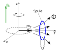
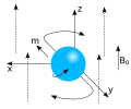
MRT Praecession Rotation.svg 600 × 500; 2 KB
-
NIMA 2A2B3A3B13.png 260 × 234; 21 KB
-
Nlinv iter.png 873 × 491; 59 KB
-
Nmr-rekonstrukcija.png 405 × 206; 5 KB
-
Nmr-slikanje.png 405 × 235; 9 KB
-
NmrrelaxM1.gif 560 × 420; 578 KB
-
NmrrelaxM2.gif 560 × 420; 608 KB
-
Olecranon.jpg 319 × 426; 42 KB
-
Olecranon.tif 319 × 426; 218 KB
-
PAPVR.gif 240 × 176; 711 KB
-
Patchy cortical restricted diffusion as seen on ADC sequence.jpg 384 × 384; 65 KB
-
Patient and MRI device.svg 532 × 435; 86 KB
-
Pauly5.jpg 3,289 × 1,649; 1.43 MB
-
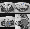
Pelvic MRI 125131.png 473 × 480; 122 KB
-
PET attenuation correction for hybrid MRI PET imaging system.png 1,444 × 1,187; 742 KB
-
PMC2730233 01-0273-F1.png 512 × 342; 170 KB
-
PMC4928621 ijcn-10-086-g002.png 512 × 683; 497 KB
-
Processo de relaxação.png 391 × 203; 7 KB
-
Proteção magnética.png 362 × 325; 13 KB
-
Radialvscart.png 424 × 863; 71 KB
-
Radiologist interpreting MRI.jpg 870 × 580; 72 KB
-
Relaxation longitudinal magnetization.svg 1,500 × 625; 109 KB
-
Relaxation transverse magnetization.svg 1,500 × 625; 93 KB
-
Researcher-test.jpg 450 × 217; 50 KB
-
RESONANCIA MAGNÉTICA DE LA ATM.jpg 1,055 × 987; 443 KB
-
Ressonància magnètica cerebral amb i sense amúsia.jpg 10,732 × 5,605; 5.15 MB
-
RF shielding waveguide for MRI.jpg 4,000 × 3,000; 4.56 MB
-
RM de abdomen con gadolinio.jpg 284 × 363; 43 KB
-
RMN exemplo.png 543 × 300; 57 KB
-
Saghead2.jpeg 224 × 224; 24 KB
-
SCC MRI.png 3,000 × 2,948; 2.62 MB
-
SE table.jpg 625 × 414; 129 KB
-
Single islet.jpeg 1,005 × 1,305; 1.06 MB
-
Spin c13.png 319 × 239; 11 KB
-
Spin Echo diagram.svg 1,250 × 875; 496 KB
-
Sprgelenkli131107.jpg 564 × 447; 37 KB
-
Stack plot serial 31P MRS calf muscle.jpg 4,284 × 2,304; 935 KB
-
Stenose-MRI.jpg 401 × 435; 67 KB
-
Sulcus centralis - Identification axial - MRI T2.jpg 681 × 795; 69 KB
-
Sulcus centralis - Identification sagittal - MRI T2.jpg 912 × 700; 66 KB
-
Suszeptibilitaetsartefakte durch festsitzende Zahnspange 13W - MR - 001.jpg 2,259 × 1,801; 333 KB
-
Suszeptibilitaetsartefakte durch festsitzende Zahnspange 13W - MR - 002.jpg 2,259 × 1,801; 344 KB
-
Sweep diagram for hyperbolic secant pulse.png 406 × 618; 35 KB
-
Syndrome de Joubert IRM PAMJ-22-127-g001.jpg 566 × 215; 43 KB
-
T1 mapping jcmr fig1.png 1,301 × 686; 69 KB
-
T1map scale.png 139 × 973; 60 KB
-
T1RelaxationMalayalam.gif 960 × 720; 140 KB
-
T2RelaxationMalayalam.gif 960 × 720; 178 KB
-
Tablainterrferencia.jpg 891 × 395; 120 KB
-
The Specialists(MRI).jpg 4,404 × 5,240; 7.12 MB
-
Thoracic duct MRL in lymphoedema.tif 1,870 × 2,079; 15 MB


_Unit%2C_Cromer_and_district_Hospital%2C_Mill_Road%2C_Cromer.JPG/120px--2018-11-18_MRI_(Magnetic_resonance_imaging)_Unit%2C_Cromer_and_district_Hospital%2C_Mill_Road%2C_Cromer.JPG)
_and_MRI_Unit%2C_Cromer_Hospital.JPG/120px--2022-01-10_The_Mammography%2C_DEXA_(osteoporosis_diagnosis)_and_MRI_Unit%2C_Cromer_Hospital.JPG)



.png/120px-872-4-8_(n).png)










_-_journal.pone.0017879.g001.png/82px-CT_and_MRI_of_a_red-eared_slider_(Trachemys_scripta)_-_journal.pone.0017879.g001.png)

_Axial_MR_image_IR.jpg/120px-Delayed_(10_min_)_Axial_MR_image_IR.jpg)




_of_a_whole_maize_plant_and_root_systems_of_a_bean_and_a_sugar_beet_plant_-_Plphys_v170_3_1176_f1.jpg/60px-Magnetic_Resonance_Images_(MRI)_of_a_whole_maize_plant_and_root_systems_of_a_bean_and_a_sugar_beet_plant_-_Plphys_v170_3_1176_f1.jpg)







.jpeg/96px-MRI_Image_of_Human_Head_(94-087-1).jpeg)









.jpg/101px-The_Specialists(MRI).jpg)


